At imed, all research groups benefit from shared scientific platforms, including core laboratories and technologies, and broad technical support, as well as administrative support services, which create a privileged environment for research, training, and outside services.
Resources & Services
Animal Facility
Head: Maria Manuela Gaspar
The Animal Facility supports the discovery and development of innovative medicines for the benefit of humans and animals. Animal experiments have been vital in pharmaceutical and biomedical research. Future advances in the treatment and diagnosis of human diseases will continue to depend on inquiries that use animals and other alternatives.
This is a conventional facility for the housing of laboratory rodents licensed by the National Authorities “Direção Geral de Alimentação e Veterinária” (DGAV). All animal experiments conducted in the Animal Facility are subject to rigorous review and must be previously submitted to the Animal Welfare Board (ORBEA – Orgão de Bem-Estar Animal) at the Faculty of Pharmacy, University of Lisbon (Regulamento 806/2016), and then approved by DGAV, the competent national authority responsible for implementing the legislation for the protection of animals for scientific purposes. Together, they ensure that research animals are used only when necessary and under humane conditions. Personnel and users are certified researchers for conducting animal experimentation and procedures are performed according to the EU Directive (2010/63/UE) and Portuguese laws (Decreto Lei 113/2013, Despacho 2880/2015 and Portaria 260/2016). iMed.ULisboa is committed to conducting the highest-quality research and to providing animals used in research with the best care available. Alternatives to animal use, which include computer modeling, cell culture and bacterial systems are often used at iMed.ULisboa.
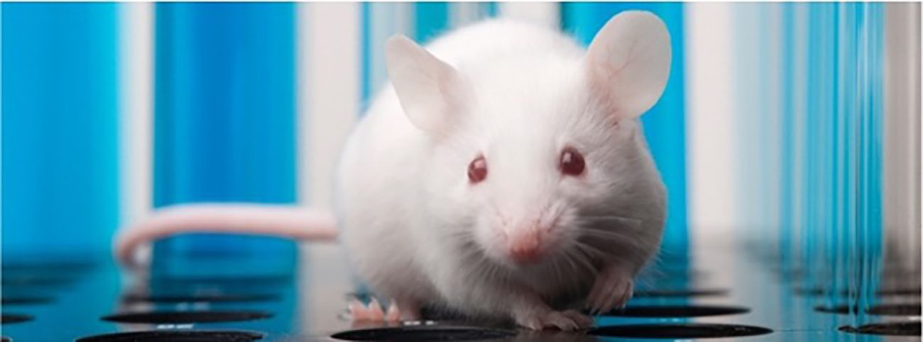
The Animal Facility provides technical and scientific support to investigators on protocol development, refinement of experimental procedures, small surgery techniques, and services of husbandry and routine daily care (feeding, watering, and cage changing). Several animal models are established and under routine use in biodistribution and toxicity studies and disease modeling studies, including animal models of infection, acute and chronic inflammation, xenograft tumors, non-alcoholic fatty liver disease, neurodegenerative diseases. Upon request and contract, these or other animal models may be provided to external entities.
The Animal Facility consists of several rooms for animal maintenance with housing capacity of around 500 small rodents (rats and mice), and rooms for experimental procedures (small surgeries and dissections). Metabolic cages are also available. Support rooms are used for cleaning, washing and sterilization of cages and other equipment, food and bedding.
Cell Culture
Head: Joana Amaral, Rui Silva
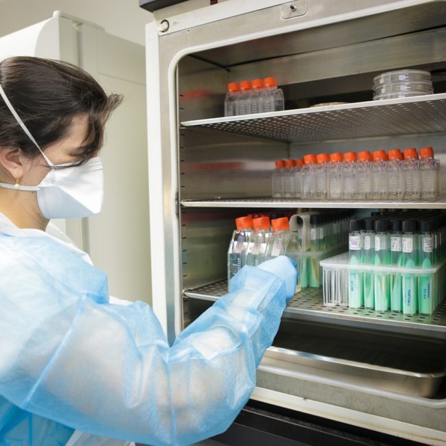
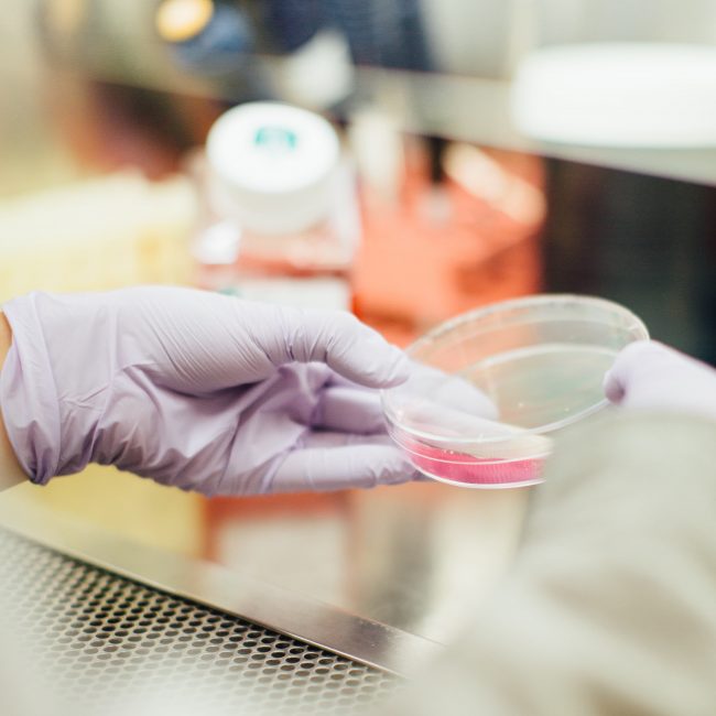
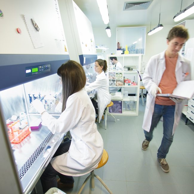
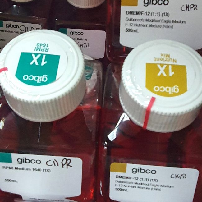
The Cell Culture Facility comprises dedicated cell culture rooms equipped with the required environment and equipment for a wide range of cell and tissue culture procedures, from maintenance and manipulation of cell lines and tissue samples, to cell observation and data analysis. In addition, the facility provides routine mycoplasma detection testing for mammalian cell lines.
Consists of laminar flow hoods (Esco, Class II Type A2), CO2 incubators (Hera Cell), inverted microscopes (Zeiss) coupled to an imaging system (Leica), and support equipment (centrifuges, water baths, refrigerators, freezers). Fluorescence and bright-field microscopes (Zeiss) with dedicated cameras (Leica) and imaging and acquisition systems are available.
Additional dedicated equipment provides cell analysis high-throughput capabilities with Multidrop Combi Reagent Dispenser (Thermo Scientific) for 6 to 1536-well plates; GloMax®-Multi+Microplate Multimode Reader (Promega), accepting 6 to 384-well plates, and accommodating luminometer, fluorescence, and visible/UV absorbance modules and dual injector system for 6 to 96-well plate formats; and xCELLigence RTCA SP (ACEA Biosciences) for real-time label free impedence-based cell analysis in 96-well format.
Provides biological evaluation of cell function, routinely determining the role of transgenes and the cytotoxic and cytoprotective activities of synthetic and natural compounds in multiple cell models, including immortalized cells (human, monkey, rat, mouse), primary cultures (rat and mouse liver, brain), and embryonic stem cells (rat and mouse).

Gene Expression
Head: Rui Castro
The Gene Expression Facility is dedicated to sample quality monitoring and analysis of gene expression at DNA, RNA and protein levels, routinely performing a multitude of biochemical and molecular biology techniques.
Consists of equipment for sample quality monitoring and gene expression analysis, including a Qubit 2.0 fluorometer (Thermo Fisher Scientific) and a NanoDrop 2000c spectrophotometer (Thermo Scientific), standard gel electrophoresis (Bio-Rad), Trans-Blot Turbo System (Bio-Rad), absorbance plate readers (Bio-Rad), Chemidoc MP and Chemidoc XRS gel/membrane and X-ray film imaging (Bio-Rad), end-point thermocyclers (Bio-Rad and Thermo Scientific), and real time PCR systems including the 7300 Real-Time PCR (Applied Biosystems) and sate-of-the-art QuantStudio™ 7 Flex Real-Time PCR System (Applied Biosystems). The later enables high-throughput, quantitative gene expression, combining 384-well microfluidic gene expression, predesigned or customed card arrays, with multiplexing (21 filter combinations), and fast real-time capabilities.
Additional equipment includes a Guava easycyte 5HT benchtop flow cytometer (Merck Millipore) for high-throughput cell analysis in 96-well format, and stopped-flow (HiTech Scientific) and patch-clamp setups (Axon Instruments) used for kinetic analyses of ion channel activity.
Provides technical support ranging from advice in experimental design to data analysis.
Radioisotope
Head: M. Luísa Corvo
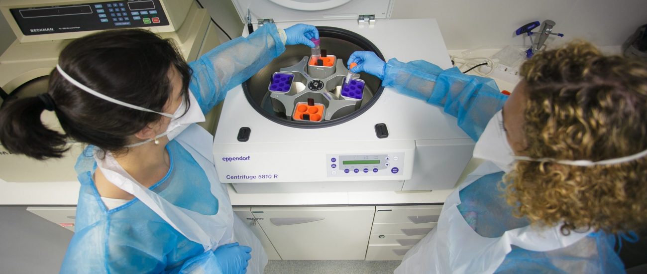
The Radioisotope Facility (gamma and beta emission) follows strict rules driven by requirements of improved safety for workers on radioactive sample handling. All new users must perform radiation safety training.
Consists of dedicated areas for labeling of proteins, other macromolecules and low molecular weight compounds by chemical modification.
Provides pharmacokinetics, biodistribution, and metabolite studies to investigators or external entities upon request and contract. The physical proximity to the Animal Facility enables in vitro and in vivo studies.

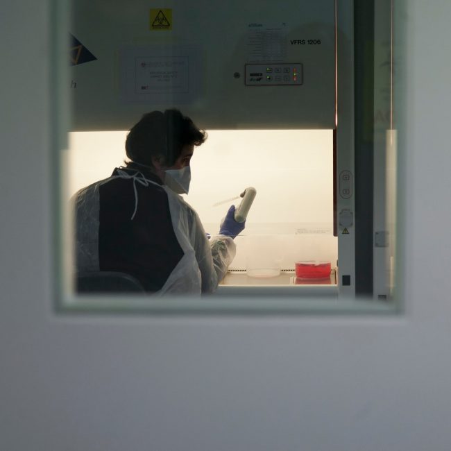
Biosafety Level 3
Head: Quirina Santos Costa
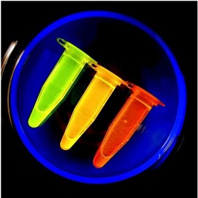
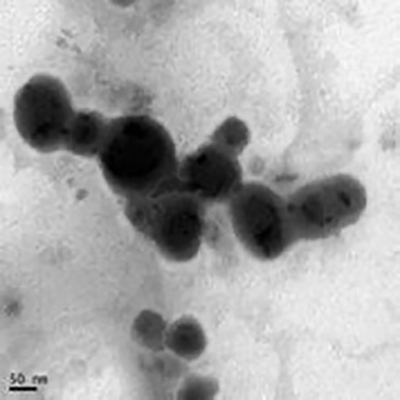
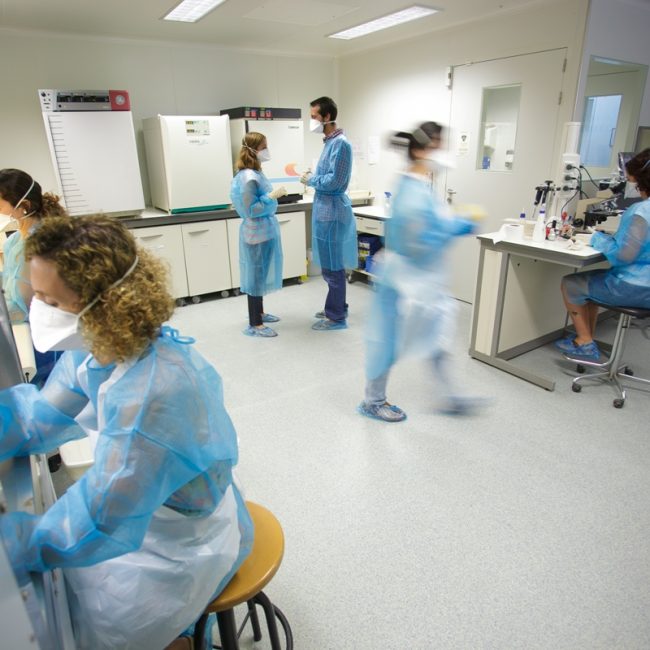
The Biosafety Level 3 Facility is specifically dedicated to research involving biological pathogens of level 3 security. It was designed to minimize the risk of personnel and environmental exposure to potential hazardous agents according to European and Portuguese legislation. All users must undergo specific biosafety level 3 training, and must follow strict rules and guidelines while working in the facility.
Consists of an anteroom for material and personnel preparation, and a main procedure room equipped with tree vertical laminar flow chambers (type A2 and type B2), three CO2 incubators (Hera Cell), one regular incubator, two benchtop centrifuges (Eppendorff), a benchtop ultracentrifuge (Beckman), an aerosol-tight microfuge (Eppendorff), a Tecan infinite 200 multimode microplate reader, water baths, freezers, refrigerators, optical and inverted phase-contrast microscopes (Leica), and a dedicated double door pass-through autoclave (Matachana).
Mass Spectrometry
Head: Maria do Rosário Bronze
The Mass Spectrometry Facility is part of the National Mass Spectrometry Network.
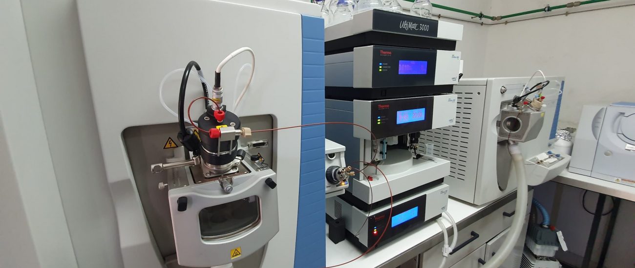
Consists of a Triple Quadruple mass spectrometer (Micromass Quattro Micro API, Waters) with electrospray ionization (ESI) atmospheric pressure chemical ionization (APCI) ion sources. This facility is also equipped with an Ion-Trap (LCQ-Fleet, Thermo) mass spectrometer dedicated to the characterization of proteins and biological conjugates.
Provides identification and quantification of small molecules in complex matrices, as biological fluids and extracts of natural products. Services are available for users on a “do-it-yourself” basis or self-service, for long-term studies, upon initial training requirements. A technician is also available for a full-service.
Molecular BioScreening
Head: Cecília Rodrigues, Vanda Marques
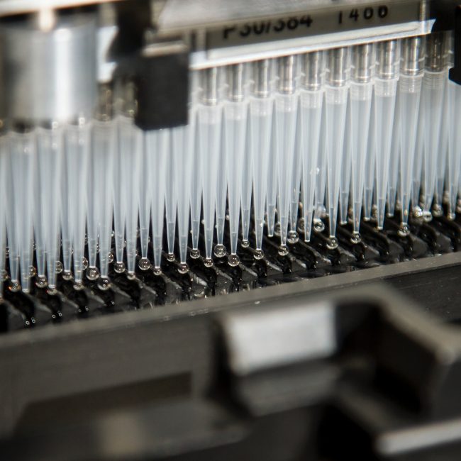
Molecular BioScreening is the newest infrastructure from imed that offers an innovative and integrated approach of cell-based medium- to high-throughput assays for screening small molecules (natural or synthetic) and biologics, ultimately leading to discovery of new therapeutics. It combines the power of relevant cell models, phenotypic screens and live cell functional assays to both model disorders and search for drugs to treat disease

BioImaging
Head: Liana Silva
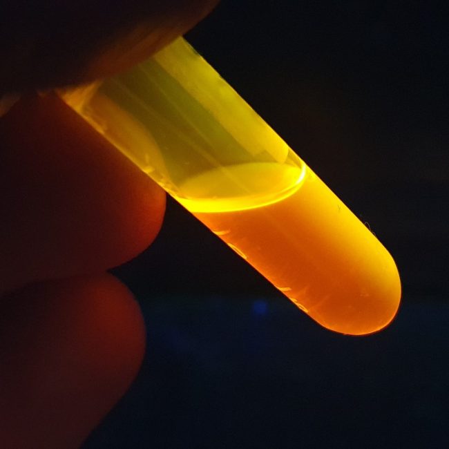
At imed there are several resources available for bioimage acquisition.
Both fluorescence and bright-field microscopes (Zeiss) with dedicated cameras (Leica) and imaging and acquisition systems are available within the Cell Culture facility.
The Leica TCS SP8 laser scanning confocal microscope is a new resource at iMed.ULisboa for fluorescence imaging. The inverted DMi8 fluorescence microscope is equipped with a fully motorized stage, fast z movement (Leica Super Z Galvo stage), 4 solid state lasers (405, 488, 552, 638 nm), four detectors (one HyD high-sensitivity, three PMT), a transmitted light detector with CCD camera, three dry objectives (5x, 10x and 20x) and two oil immersion objectives (40x and 63x), and allows several types of image acquisition (2D, z-stack and time-lapse).

Nuclear Magnetic Resonance
Head: Noélia Duarte
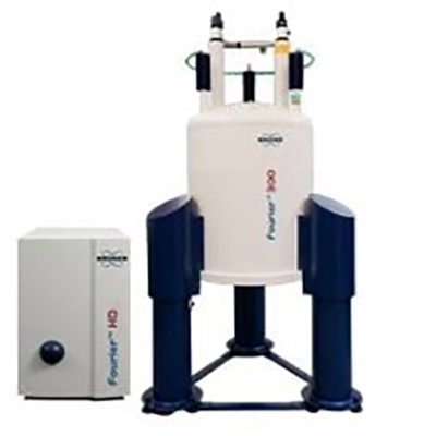
The Nuclear Magnetic Resonance (NMR) Facility is equipped with a Bruker Fourier 300 MHz Spectrometer.
Promotes the use of nuclear magnetic resonance spectroscopy in the areas of structure-based drug design, including structure characterization and fragment-based drug design.


Computer Assisted Drug Design
Head: Rita Guedes
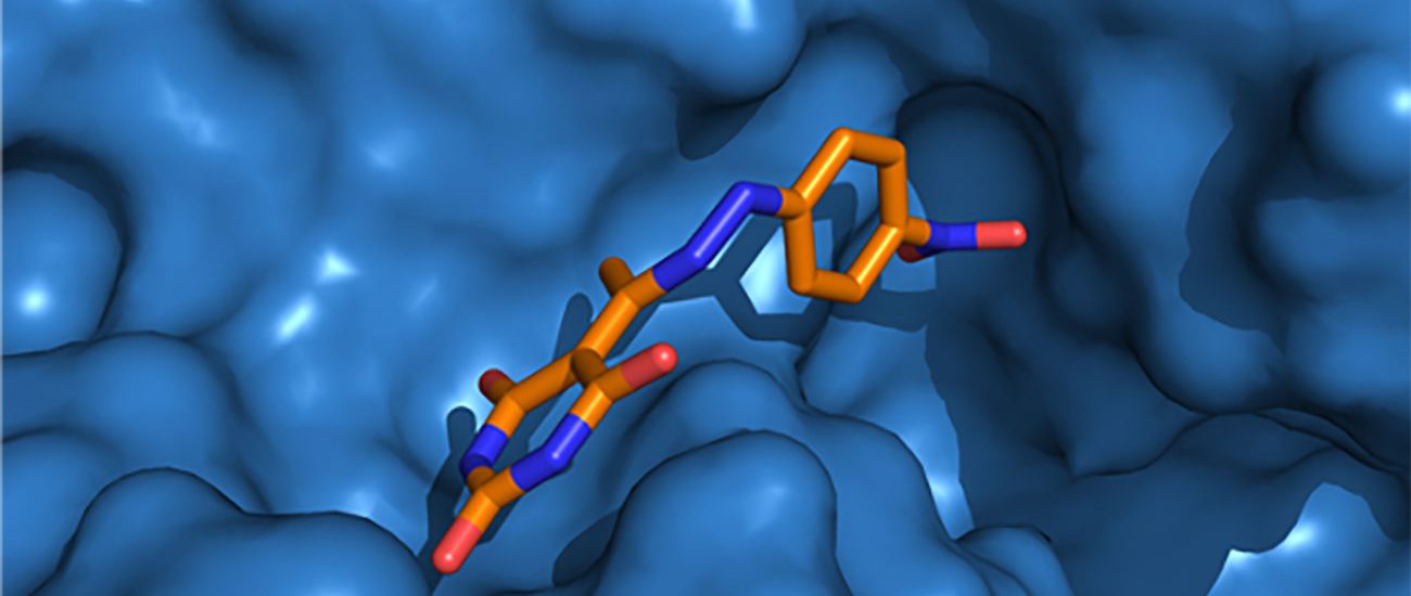
The Computer Assisted Drug Design Facility consists of a Linux-based high performance computer cluster with 424 CPU cores, 4 to 8GB per CPU/GPU and 2 TB per node with a specific implementation of state-of-the-art software for molecular modeling, molecular dynamics, virtual screening, and de novo design.
Provides technical support ranging from advice in experimental design to data analysis.
Flow Cytometry
Head: Catarina Godinho Santos; Staff: Miguel Cardoso
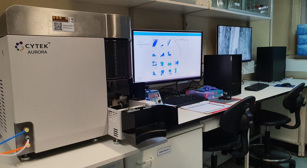
The Aurora system at iMed.ULisboa consists of the Cytek® Aurora full spectrum flow cytometer and a computer workstation running SpectroFlo® software for sample acquisition and data analysis. This spectral flow cytometry system allows unique fluorochrome combinations in comparison to conventional flow cytometry and enables analysis of cells with high autofluorescence.
The cytometer is an air-cooled, compact benchtop instrument. It is equipped with 4 lasers (Violet, Blue, Yellow-Green and Red), 48 detection channels for fluorescence, and three channels for scatter (blue laser FSC, blue laser SSC, and violet laser SSC). Sheath and waste fluids are contained in 4-L tanks, included with the system. High-throughput sample loaders are available to automate sample delivery and acquisition and currently are compatible with 96-well plates.
An independent workstation for analysis of flow cytometry data is available upon booking, where SpectroFlo®, FCS ExpressTM 7 and FlowJoTM software can be used.
Technical support ranging from advice in experimental design to data analysis can be requested.
Available for iMed.ULisboa and external researchers.
